Addressing Dorsal Metatarsal Cuneiform Exostosis: Treatment Approaches for Saddle Bone Deformity
Dorsal metatarsal cuneiform exostosis, commonly known as saddle bone deformity, represents a challenging orthopedic condition affecting the midfoot. This bony prominence develops on the dorsal (top) surface of the foot where the metatarsal bones meet the cuneiform bones, creating a painful bump that can significantly impact daily activities and quality of life. Understanding the various treatment modalities available, from conservative management to surgical intervention, is essential for patients and healthcare providers navigating this condition.
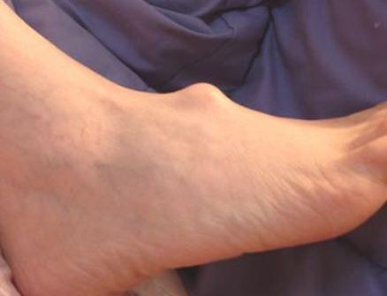
The saddle bone deformity typically manifests as a visible and palpable bony prominence on the top of the foot, often causing discomfort when wearing shoes, particularly those with rigid uppers or laces that cross directly over the affected area. The condition may arise from various factors, including genetic predisposition, biomechanical abnormalities, trauma, or repetitive stress. Some individuals are born with a natural prominence in this area, while others develop the exostosis over time due to degenerative changes or inflammatory conditions such as osteoarthritis.
Conservative management represents the first line of treatment for most patients with dorsal metatarsal cuneiform exostosis. The primary goal of non-surgical approaches is to reduce pressure on the bony prominence, minimize inflammation, and alleviate pain. Footwear modification stands as one of the most effective conservative strategies. Patients should seek shoes with a high, wide toe box that provides adequate vertical clearance over the midfoot region. Soft, flexible uppers made from leather or stretch materials accommodate the prominence better than rigid synthetic materials. Avoiding shoes with laces or straps that cross directly over the affected area can dramatically reduce irritation and discomfort.
Orthotic devices play a crucial role in conservative management by addressing underlying biomechanical issues that may contribute to the condition. Custom or over-the-counter arch supports can help redistribute pressure away from the painful prominence and improve overall foot alignment. Some orthotists create specialized pads or cutouts that surround the exostosis, effectively creating a protective "doughnut" that shields the area from direct pressure while supporting the surrounding structures.
Padding and protective devices offer immediate symptomatic relief for many patients. Gel pads, moleskin, or specialized cushioning products can be applied directly over or around the bony prominence to create a buffer between the foot and shoe. These simple interventions often provide sufficient comfort for individuals with mild to moderate symptoms, particularly when combined with appropriate footwear.
Anti-inflammatory medications, both topical and oral, help manage pain and reduce inflammation associated with the condition. Nonsteroidal anti-inflammatory drugs (NSAIDs) such as ibuprofen or naproxen can be used during acute flare-ups, while topical preparations may provide localized relief without systemic effects. Ice application after activities that aggravate the condition can further reduce inflammation and discomfort.
Physical therapy interventions may benefit some patients by addressing muscle imbalances, improving joint mobility, and teaching techniques to reduce stress on the affected area. Stretching exercises for the Achilles tendon and calf muscles can help optimize foot biomechanics, while strengthening exercises for the intrinsic foot muscles may improve overall foot stability and function.
When conservative measures fail to provide adequate relief after several months of consistent implementation, or when the deformity is severe enough to significantly limit function despite optimal non-surgical management, surgical intervention may be warranted. The decision to proceed with surgery should involve careful consideration of the patient's symptoms, functional limitations, overall health status, and expectations.
The surgical procedure for dorsal metatarsal cuneiform exostosis, known as exostectomy or cheilectomy, involves removing the excess bony prominence to create a smoother contour on the top of the foot. The surgery is typically performed under regional anesthesia on an outpatient basis. The surgeon makes an incision over the affected area, carefully dissects through the soft tissues, and uses specialized instruments to resect the bony prominence. The amount of bone removed depends on the size of the exostosis and the underlying joint condition. Care must be taken to remove sufficient bone to alleviate symptoms while preserving joint stability and function.
In cases where significant arthritis accompanies the bony prominence, the surgeon may need to perform additional procedures such as joint debridement or, in severe cases, arthrodesis (fusion) of the affected joints. These more extensive procedures carry longer recovery times but may be necessary for optimal long-term outcomes in patients with advanced degenerative changes.
Post-operative recovery typically involves a period of protected weight-bearing, often using a surgical shoe or walking boot. Most patients can bear weight on the foot within a few days to weeks, depending on the extent of the procedure. Physical therapy during the recovery phase helps restore range of motion, strength, and normal gait patterns. Swelling may persist for several months after surgery, and patients should maintain elevation and compression as recommended by their surgeon.
Surgical outcomes for dorsal metatarsal cuneiform exostosis are generally favorable, with most patients experiencing significant pain relief and improved shoe tolerance. However, as with any surgical procedure, potential complications exist, including infection, nerve damage, recurrence of the prominence, and continued pain. Careful patient selection, meticulous surgical technique, and appropriate post-operative management help minimize these risks.
Prevention strategies for individuals at risk of developing saddle bone deformity include wearing properly fitted footwear with adequate midfoot clearance, maintaining healthy body weight to reduce mechanical stress on the feet, and addressing biomechanical abnormalities through orthotic support. Early intervention at the first signs of discomfort may prevent progression of the condition.
Fixing a saddle bone deformity requires a comprehensive, individualized approach that begins with conservative management and progresses to surgical intervention only when necessary. The combination of appropriate footwear, protective padding, orthotic support, and anti-inflammatory measures successfully manages symptoms for many patients. For those who remain symptomatic despite optimal conservative care, surgical exostectomy offers excellent pain relief and functional improvement, allowing return to normal activities and comfortable shoe wear.
Post Your Ad Here
Comments