The Diagnosis of Chilblains in the Feet
Chilblains, medically known as pernio or perniosis, represent a localized inflammatory condition affecting the small blood vessels in the skin, most commonly occurring in the feet and toes. While relatively uncommon in temperate climates, chilblains remain a significant dermatological concern in cold, damp environments, affecting individuals who experience repeated exposure to cold without adequate protection. Understanding the diagnostic approach to chilblains is essential for healthcare providers, as the condition can be confused with other dermatological and vascular disorders, and proper identification ensures appropriate management and prevention of recurrence.
Clinical Presentation and Recognition
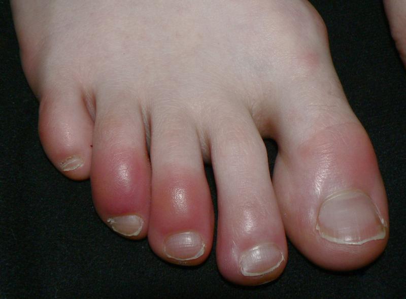
The diagnosis of chilblains begins with recognition of its characteristic clinical features. Patients typically present with erythematous or violaceous lesions on the dorsal surfaces of the toes, though the heels, lateral aspects of the feet, and other exposed areas may also be affected. These lesions manifest as inflammatory papules, nodules, or plaques that develop several hours to days following cold exposure. The affected areas often appear swollen and may have a dusky, bluish-purple discoloration that distinguishes them from simple cold injury.
Patients commonly report intense itching and burning sensations in the affected areas, which can be quite distressing and may interfere with daily activities and sleep. In more severe cases, blistering, ulceration, or even tissue necrosis can occur, though this is relatively uncommon. The symptoms typically worsen upon rewarming, a characteristic feature that helps distinguish chilblains from other cold-related injuries. This paradoxical worsening occurs because rapid rewarming causes reactive hyperemia and inflammation in the damaged tissue.
Patient History and Risk Factors
A thorough patient history is crucial in diagnosing chilblains. Clinicians should inquire about recent cold exposure, particularly prolonged exposure to cold, damp conditions without adequate foot protection. The condition is more prevalent during winter months and in individuals living in homes with inadequate heating. However, chilblains can also occur during transitional seasons when temperature fluctuations are common.
Several demographic and physiological factors increase susceptibility to chilblains. Women are affected more frequently than men, and the condition shows a predilection for young adults, though it can occur at any age. Individuals with low body mass index appear to be at higher risk, possibly due to reduced subcutaneous fat providing less insulation. A personal or family history of chilblains is significant, as there appears to be a hereditary component with some individuals demonstrating abnormal vascular reactivity to cold.
Certain medical conditions predispose individuals to chilblains. Raynaud's phenomenon, characterized by exaggerated vasospastic responses to cold, frequently coexists with chilblains. Connective tissue diseases, particularly systemic lupus erythematosus, can present with chilblain-like lesions. Smoking is a notable risk factor, as nicotine causes vasoconstriction and impairs peripheral circulation. Additionally, medications that affect peripheral circulation, such as beta-blockers, may increase susceptibility.
Physical Examination
The physical examination focuses on careful inspection of the feet and assessment of peripheral circulation. The distribution of lesions is diagnostically important—chilblains typically affect the toes symmetrically, though asymmetric presentation can occur. The examiner should note the color, size, and configuration of lesions, as well as the presence of any secondary changes such as excoriation from scratching, blistering, or ulceration.
Palpation of the lesions typically reveals them to be tender, and the surrounding skin may feel cool to touch initially, though temperature normalizes upon rewarming. Assessment of peripheral pulses is essential to rule out underlying vascular insufficiency that might predispose to more serious cold injuries. The dorsalis pedis and posterior tibial pulses should be palpated and documented. Capillary refill time can provide additional information about peripheral perfusion.
The examination should extend beyond the feet to identify any systemic features that might suggest an underlying condition. Signs of connective tissue disease, such as malar rash, sclerodactyly, or digital ulcers, should be sought. The presence of livedo reticularis might suggest a vasculopathy or autoimmune condition.
Differential Diagnosis
Distinguishing chilblains from other conditions is a critical diagnostic challenge. Frostbite represents a more severe cold injury involving actual tissue freezing and typically occurs at much lower temperatures than those that cause chilblains. Frostbite lesions are usually more severe, with clear demarcation between viable and non-viable tissue in advanced cases.
Vasculitis can present with similar purpuric lesions on the feet but typically involves palpable purpura and may be associated with systemic symptoms. Erythromelalgia causes burning pain and erythema in the feet but is typically triggered by warmth rather than cold. Acrocyanosis produces persistent bluish discoloration of the extremities but lacks the inflammatory papules characteristic of chilblains.
In recent years, chilblain-like lesions gained prominence as a manifestation of COVID-19, particularly in young adults and children. These "COVID toes" presented diagnostic challenges and highlighted the importance of considering current infectious disease trends when evaluating similar lesions.
Diagnostic Investigations
Chilblains remains primarily a clinical diagnosis based on history and physical examination. However, certain investigations may be warranted in specific circumstances. Routine laboratory investigations are generally unnecessary for straightforward cases with clear cold exposure history and typical presentation.
When systemic disease is suspected, serological testing may be indicated. Autoimmune screening, including antinuclear antibodies, rheumatoid factor, and complement levels, can help identify underlying connective tissue disorders. Cryoglobulin testing may be considered if cryoglobulinemia is suspected.
Skin biopsy is reserved for atypical cases or when the diagnosis remains uncertain despite clinical evaluation. Histopathological examination typically reveals superficial and deep perivascular lymphocytic infiltrate with papillary dermal edema. These findings, while supportive, are not entirely specific to chilblains.
The diagnosis of chilblains in the feet relies heavily on clinical acumen, combining careful attention to the characteristic presentation with thorough history taking regarding cold exposure and risk factors. While typically a benign condition, proper diagnosis ensures appropriate patient education about prevention and distinguishes chilblains from more serious vascular or systemic conditions requiring different management approaches.
Post Your Ad Here
Comments