Role of Pelvic Ultrasound in Pregnancy
A pelvic ultrasound can give complete information about your reproductive
organs and a pregnancy. It might be utilized amid fertility treatments too.
Being non-invasive, exact, fairly low risk and affordable, it has turned into indispensible
tool in observing pregnancy, particularly when high risk.
What is a
pelvic ultrasound?
The main purpose of pelvic ultrasound is amid pregnancy. At the time of
this procedure, high-frequency sound waves develop images of the pelvic organs.
The sound waves entered into pelvis, and measure how they reflect – or echo –
back from the distinct tissues. Also, it can find fibroid tumors, cysts,
growths, and issues with fallopian tubes. There are two ways to perform this
test.
·
Transabdominal
ultrasound. In this way, a small handheld device known as transducer is passed to
and fro over the lower belly to look for large uterine fibroids or other
problems.
·
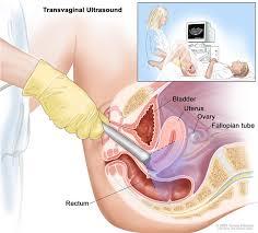
Transvaginal
ultrasound. In this method, the transducers us made to fit into a woman’s vagina. A
few women may have both transabdominal as well as TVS ultrasound to look at
the complete pelvic area. A hysterosonogram (in rare cases) may also be done to
check inside of the uterus. At times, a tiny sample of tissue (biopsy) may be
taken with little tools inserted through the vagina amid a transvaginal
ultrasound.
When might you
be asked to do a pelvic ultrasound?
·
Early
pregnancy: When it is your 5-7 weeks of pregnancy, the ultrasound may be done to
determine the baby size to confirm the supposed due date, find multiple babies,
or confirm that the babyis alive (viable).
·
Ectopic
pregnancy: Additionally, it is mainly helpful in differentiating between
intrauterine (within the uterus) as well as ectopic (outside the uterus)
pregnancies.
·
Middle of the
pregnancy: When you are about 16–20 weeks, the process can confirm baby growth, show
defects in the anatomy of the baby, and detect the amniotic fluid and placenta.
·
Toward the end
of pregnancy: It may be utilized to access fetal size, growth, position,
or to check the placenta.
·
Prenatal
genetic testing: Doctors may utilize ultrasound to direct the biopsy
needle at the time of chorionic villus and amniocentesis sampling. The imaging permits
placement of the long needle that is put into the patient's uterus to gather
cells from the placenta or amniotic fluid.
·
Early labor: May be it is
utilized to check the cervical length in pregnant women who are at risk for
preterm labor.
A few other reasons you may be asked to do a pelvic ultrasound:
·
To check what is causing pelvic pain
·
Identify the reason behind vaginal bleeding
·
Look for pelvic inflammatory disease (PID)
·
Find an intrauterine device (IUD).
·
Check the shape and size of the uterus and the
thickness of the uterine lining (endometrium)
·
Look at the size and shape of the ovaries
·
Determine the size and condition of the ovaries at the
time of infertility treatment
·
Determine a lump found amid a pelvic examination
·
Determine uterine fibroids (and their growth) found amid
a pelvic examination
Hence, this type of Ultrasound
in Gurgaon is highly advised by the doctors to determine certain things
which are mentioned above. Just make sure you visited the right center for an
ultrasound.
Post Your Ad Here
Comments