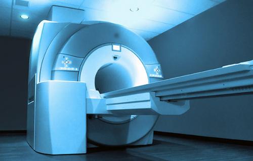
WHAT THE MRI SHOWS

Magnetic resonance imaging visualizes soft tissue. It is used to study the brain, internal organs, joints, muscles, subcutaneous fatty tissue, and blood vessels. MR imaging is a good complement to X-ray or CT scanning of the spine, as it clearly visualizes discs, nerve roots, spinal canal, and other soft tissue structures.
The method is often used in controversial and complex clinical situations. By focusing on what the MRI shows, doctors are able to make the right diagnosis and choose the optimal therapeutic tactics. Magnetic resonance imaging is recognized as a harmless and safe procedure that is approved for children and pregnant women.
What a CT scan reveals depends on the purpose and type of study. A native scan is prescribed to assess the structure of organs, MR angiography is used to study blood vessels, and contrast enhancement is performed if inflammation and tumors are suspected.
What diseases can MRI detect?
Magnetic resonance imaging takes layer-by-layer pictures of the area under study. The slices are made in 1 mm increments, in three mutually perpendicular planes. The images alternate between light and dark areas, whose contours correspond to tissues with different fluid content.
MRI visualizes internal structures. The procedure allows you to assess the anatomy of the body and tissue structure. Deviations from the norm may indicate individual features of the body, developmental abnormalities, trauma, or disease progression.
Pathological processes are detected by specific signs - changes in signal intensity, mass effect, accumulation of contrast agent, etc.
Without medical education, experience and skills in the field of radiation diagnostics, it is virtually impossible to understand what an MRI shows. A radiologist is responsible for deciphering tomograms. After examining the scans, the specialist logs the changes detected. In some centers, the doctor briefly explains the detected abnormalities when giving the results to the patient. The clinician uses the MRI report to make a diagnosis, select treatment, monitor the dynamics of the disease, and evaluate the effectiveness of therapy.
The MRI Orlando shows signs of:
- abnormalities of organ development;
- benign masses;
- malignization and the presence of metastases;
- inflammatory processes;
- pus accumulation (abscess, phlegmon);
- replacement of functionally active tissue with connective fibers;
- Ischemia and necrosis;
- degenerative processes, etc.
The method is indispensable in neurology and neurosurgery for detection of brain or spinal cord pathologies. The study of blood vessels reveals anomalies in the structure of the bloodstream, stenosis, aneurysms, thrombosis, atherosclerosis, wall dissection, vasculitis, etc.
What won't the MRI be able to show?
Magnetic resonance imaging will be of little use in diagnosing functional disorders that have no organic basis (some hormonal failures, autonomic reactions, etc.). The procedure is insufficiently informative regarding bone diseases. MRI can show indirect signs of changes in dense structures, but it is difficult to detect small defects.
MR angiography of arteries and veins is appropriate for suspected diseases that are not accompanied by vascular wall damage. CT is more appropriate for diagnosing acute cerebral circulation disorders.
When prescribing the procedure, it is important to adequately assess the appropriateness of the procedure. Magnetic resonance imaging of the lungs is not informative, since the latter have a loose structure and are filled with air. MR imaging of hollow and moving organs (stomach, intestines, etc.) requires specific preparation and does not always allow to display objects of interest in detail. Images of the abdominal cavity will show large tumors, extensive inflammation. Small neoplasms may go unnoticed due to artifacts caused by intestinal peristalsis.
Whether MRI detects pathology also depends on the power of the equipment. Older models of CT scanners do not provide clear and detailed images. Closed high-field machines can produce more detailed images.
The diagnostic value of the results depends on the professionalism of the radiologist. If the doctor lacks experience, he or she may not notice the signs of diseases or may interpret the changes incorrectly. It is impossible to exclude the possibility of 100% error. In order to reduce the risk of false results, it is better to perform the diagnostics in specialized centers. If the outcome of the procedure is questionable, it makes sense to obtain the opinion of another independent expert.
Advertise on APSense
This advertising space is available.
Post Your Ad Here
Post Your Ad Here
Comments