The human body contains more than 650 individual muscles
The human body contains more than 650 individual muscles
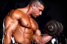
The human body contains more than 650 individual muscles attached to the skeleton, which provided the impetus for movement. These muscles constitute about 40% of total body weight. The junction point of the muscle to bone or other muscles is called the source or insertion. The origin point is the junction point in fixing the muscle to bone. The insertion point is the point of attachment to the bone to the moving muscle. Generally, the muscles are attached by tough fibrous structures called tendons. These joints connect one or more joints, and the result of muscular contraction is the movement of joints. The body moves mainly by muscle groups, not individual muscles. These muscle groups pushing all kinds of actions, from threading a needle to lift heavy objects.
Adductor longus:
Adductor longus: Combined with the extensor pollicis brevis, the adductor longus muscle creates a narrow, triangular shape that surrounds the lower end of the radius (forearm bone on the thumb side). The adductor longus was born in the back of the ulna and the radius and inserts into the base of the metacarpal bone of the thumb near the palm. This Somanabolic Muscle Maximizer Extends the thumb away from the hand (ie, performs an abduction). It also rotates and flexes the hand at the wrist. The combination of the adductor longus and extensor pollicis form the group of oblique muscles of the hand, which produces a small but significant convexity in the third quarter along the bottom profile (radial) of the forearm.
Adductor longus:
Adductor longus: There are three adductor muscles in the legs, adductor longus, adductor brevis and adductor magnus. The three adductor muscles work with the pectineus to move the thigh inward. They are powerful muscles that rotate the thigh outward and move toward the opposite side, as the movement made by crossing the legs. The adductor longus is a long triangular muscle, which has its origin in both fleshy fibers as a strong tendon in a small area of the front of the pubic bone of the pelvis and inserts into the femur (upper leg bone). The adductor brevis is located behind the adductor longus. The adductor magnus is a large triangular muscle which forms a dividing wall between the muscles of the inner thigh and the back. Is located in the inner thigh. This long muscle arises from a narrow point of the pelvis, passes between the muscle masses of the popliteal tendon and quadriceps and ends at its widest appendix in the back of the femur. It is a powerful muscle that adducts the thigh. The small upper portion of the adductor magnus adductor brevis is called.
Brachial Biceps:
Brachial Biceps: The biceps brachii (arm muscle with two parts) consists of the long head and short head. It extends from the shoulder to the elbow and is the main flexor of the elbow joint. Working together with other adjacent muscles can also move the shoulder, since its upper ends are attached to the scapula (Shoulder blade). You can also rotate the lower arm so the palm faces upward, a movement called supination. At its lower end, the biceps tendon in a plane close and strong that is firmly fixed to a protrusion of the upper end of the radius. The biceps and triceps work together to control the movement up and down the forearm.
Supinator:
Supinator: The brachioradialis or supinator originates two-thirds of the length of the humerus (the bone of the upper arm) between the triceps and the brachialis. The muscle begins wide and flat and rotating to the front of the arm down. At that point it becomes to make broad and flat before ending in a flat tendon, which attaches to the radio for the thumb side. Unlike most of the forearm long tendons, tendon does not cross the wrist joint, but terminates at the distal radius. This muscle bends the arm at the elbow, although not involved in the movement of rotation of the forearm.
Deltoid:
Deltoid: The deltoid muscle is a powerful, big and thick. It has a triangular shape and a coarse texture. In its widest begins in the clavicle and the spine of the scapula (shoulder blade), the outer covering of the shoulder joint, giving the shoulder its rounded appearance, and inserted into the humerus (bone of the upper arm). This muscle moves the humerus and is used to lift the arm out from the side. Work with the pectoralis major to move the arm forward and the teres major and latissimus dorsi to move the arm back.
External oblique:
External oblique: The external oblique muscle is a large, thin sheet that covers the side of the torso and partly the front. This muscle is divided into two portions, an upper thoracic portion and a lower side portion. The thoracic portion is located along the rib cage. When the muscle is relaxed individual ribs can be seen below. The lower flank portion is located along the side of the abdomen between the ribcage and pelvis. Most of this muscle is concealed by a layer of fat. The two portions are joined at the waist. This muscle is used to bend the body forward and twist side to side.
Twins:
Twins: The calf muscles are connected to two joints, knee and ankle. They consist of a twin external, one internal and one tendon insertion. Each column is a thick muscle, separated by the back of the knee. Descending meet. The medial is larger and wraps the leg towards the front of the twin external. Both ends in the middle of the leg or slightly above, where they bind to the tendon. The twins are the fusiform bulge of the calf of the leg.
The tendon descends and fuses with the tendon of soleus, which lies just below, forming the Achilles tendon, which inserts into the heel bone. The calf muscles are pushing the body when walking, running or jumping. Raise the heel, which lifts the body. Also contributes, although minimally, to flex the knee joint.
Occipitofrontal:
Occipitofrontal: The occipitofrontal muscle is a broad-fibrous layer covering the epicranium (the top of the skull). It consists of two thin layers of muscle. The occipital portion, sometimes called the occipitalis muscle, is quadrilateral in shape and about four inches long, and covers the back of the skull. The front portion also has a quadrilateral shape. Is wider and its fibers are of greater length. Cover the front.
The frontal and occipital portions of the muscle are joined by a thin, flat tendon aponeurosis called epicranial. The fascia is placed over the muscle and covers the top of the skull. Work with the occipitofrontal muscle to move the scalp. The frontalis muscle elevates the eyebrows and scalp moves forward. The muscle occipital scalp moves backwards
Advertise on APSense
This advertising space is available.
Post Your Ad Here
Post Your Ad Here
Comments