What Does the Radiology Division Equipment Comprise?
The radiology division in a hospital is accountable for performing diagnostic imaging examinations and procedures. This may comprise X-rays, computed tomography (CT) examinations, magnetic resonance imaging (MRI) scans, and ultrasounds.
The section also completes interventional procedures, such as biopsies and angiograms. Radiologists are doctors who concentrate on understanding pictures, and they work closely with other members of the hospital staff to ensure that patients receive the best possible care.
The radiology division plays a vital role in identifying and treating many medical conditions.
What is Radiology Division Equipment?
In any hospital, you're likely to find a diversity of radiology apparatus. This comprises X-ray machines, MRI machines, CT scanners, ultrasound machines, and x-ray viewers made by X-Ray Viewer Manufacturers. This radiology equipment is vital for identifying and treating many different medical disorders.
Ultrasound Machine
An ultrasound machine is an investigative tool that uses high-frequency sound waves to create pictures of the inside of the body. The waves are directed through a transducer positioned against the skin. The echoes are then transformed into electrical impulses shown on a screen as images. Ultrasound can envisage many diverse structures in the body, including the heart, blood vessels, kidneys, and liver. It is also regularly used during pregnancy to evaluate the fetus's health. Ultrasound is harmless, effortless, and does not use ionizing energy, making it an ideal tool for diagnostic imaging.
Floor-Mounted Digital Radiography(DR)
The Floor Mounted Digital Radiography System is a digital X-ray apparatus that is suitable and familiar. It can be used to deliver general radiography amenities in most practices. It permits doctors to have imaging flexibility, image precision, and fast results in tabletop and standing examinations to make better decisions faster.
Portable Digital X-ray
A Portable Digital X-ray machine is just like a steady x-ray machine, except it is minor enough to carry around. They are used to take images of the inside of the body and can be used to identify problems with the bones, organs, and tissues.
Movable digital x-ray machines use a specific kind of camera named a charge-coupled device (CCD), which can take high-quality images. The images are then deposited on a computer and can be seen on a monitor or printed out.
Portable digital x-ray machines are becoming progressively prevalent as they offer several advantages over traditional x-ray machines. They are less costly and can be easily stirred from one location to another. In addition, portable digital x-ray machines release much lower radioactivity levels than traditional x-ray machines, making them safer for both patients and medical staff.
CT Scanner
A CT scanner is a superior type of x-ray machine. The patient lies on a table during a CT examination and is enthused by a doughnut-shaped machine. The machine takes pictures of the body from different angles as it alternates around it. The complete method typically takes less than 30 minutes.
The machine uses x-rays to take many images, or portions, of the inside of the body. It then makes a thorough image of the part being studied. The picture may be looked at on a computer monitor or printed on film.
CT examinations are effortless, fast, and provide more information than regular x-rays.
Magnetic Resonance Imaging (MRI) Apparatuses
Magnetic resonance imaging or MRI apparatuses are huge machines that use powerful magnets and radio waves to create detailed pictures of the inside of the body.
MRI machines are huge, donut-shaped machines used to identify various conditions, from tumors to torn ligaments. MRI machines are worked by radiologists, who review the pictures to look for abnormalities.
The machine itself is gaudy, and patients often wear earplugs or headphones to minimize the noise. The examination can take up to an hour, during which patients must remain motionless to avoid blurring the images.
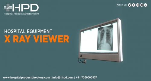
X-ray Viewer
An X-ray Viewer supplied by the X-ray Viewer Suppliers is a machine that doctors use to view x-rays and other medical pictures. Unlike old-style x-ray film, which must be advanced and viewed under a lightbox, medical pictures can be viewed immediately on a computer monitor. This permits surgeons to make identifies more quickly and accurately.
In addition, X-ray Viewers bought from X-ray Viewer Dealers often come armed with unique features that will permit doctors to zoom in on specific areas of interest or to view the pictures in three dimensions. As a consequence, X-ray viewers are an indispensable tool for identifying and treating many medical conditions.
The X-ray viewer has a lightbox that produces a cheerful, even light, making it calmer to see details in the images. The lightbox is usually installed on a stand to be located at the correct height for viewing. Additionally, the X-ray viewer typically has an exaggeration feature to see small particulars more clearly. This is mainly significant for diagnostic purposes. By consuming an X-ray Viewer, healthcare professionals can better comprehend what is going on inside a patient's body and make more precise diagnoses.
Post Your Ad Here
Comments