How Does a Neurosurgery Microscope Function?
What Is a Neurosurgery Microscope?
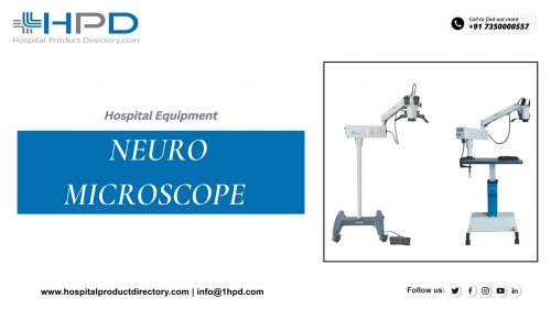
Being an essential instrument in the medical profession, the Neurosurgical microscope is used to get an enlarged image of minor objects. It reveals particulars of the assembly that are not divergent otherwise. Neurosurgery microscope made by Neuro Microscope Manufacturers plays a noteworthy role in operations involving the spinal cord, spine, and brain. It enlarges and brightens deeper parts of the operating field with utmost competence. The surgeon has a high degree of control and security due to perfect conception during the complete surgical procedure. The contemporary surgical microscope can be positioned on a stand or a table or even worn on the surgeon’s head. It is suspended on the roof or wall as well.
Operating microscopes have enhanced greatly since they were first presented. More state-of-the-art devices have also arrived in the human neurosurgical theatre, which has good exaggeration, good light without significant deviation or production of excessive warmth, and great internal constancy, which allows operational flexibility.
First and leading, although the learning curve related to the use of the microscope has been sharp and needs time, it is more than just using endoscopic-related exaggeration tools, because there is straight graphic control of the instrumentation.
The option to have exaggerations up to x10, with good complexity of the field, permits a more natural three-dimensional vision. Likened to loupes where the exaggeration and the occupied distance are fixed, surgical microscopes permit multiple different exaggerations, upholding continuous working distances and in turn, leading to outstanding suppleness and adaptability during surgical procedures.
During neurosurgery, for instance, low exaggeration is used during the boring of the vertebral lamina or the skull to safeguard that the whole surgical field is clean before sewing the muscle layers.
How Does It Work?
The extensively standard function of a microscope is the expansion of objects in the operating field. The eventual exaggeration received through a microscope is connected to the magnification lens and even the exaggeration of ocular pieces by visual principles. In a few actions, exaggeration and field depth is of main consideration. On the other side, some Neurosurgical processes which may take place at the foot of the brain, need well-lit Binocular vision in the nooks of the field. Here, the Stereoscopic Viewpoint is a highly valuable function. A Beam Splitter, an ocular device that splits an incident light ray into two or more beams, guides the imaging beam to the eyepiece and even to the camera at the same time. The light system plays a significant role too. The light strength is a noteworthy aspect of gaining graphic resolutions under the microscope made by Neuro Microscope Manufacturers.
One of the primary obstructions experienced by surgeons is physically changing the position of the microscope. A diversity of approaches concerning microscope stands were assessed and a stand is advanced in which the microscope and its fittings can be balanced by an adjustable counterweight. Even more stimulating features are video systems and certification. They are combined into the microscope system to allow the entire team to follow the procedure on a video monitor; this certainly has a positive effect on the surgery. Furthermore, video categorizations and still images can be used for exhibitions and even certification for research.
Thus, the Neurosurgery microscope is irreplaceable medical equipment that has paved the way for successful surgeries over the years.

Post Your Ad Here
Comments